Fluorescein TSA Fluorescence System Kit
Catalog No.
K1050
Fluorescein TSA Fluorescence Kit

Featured Products

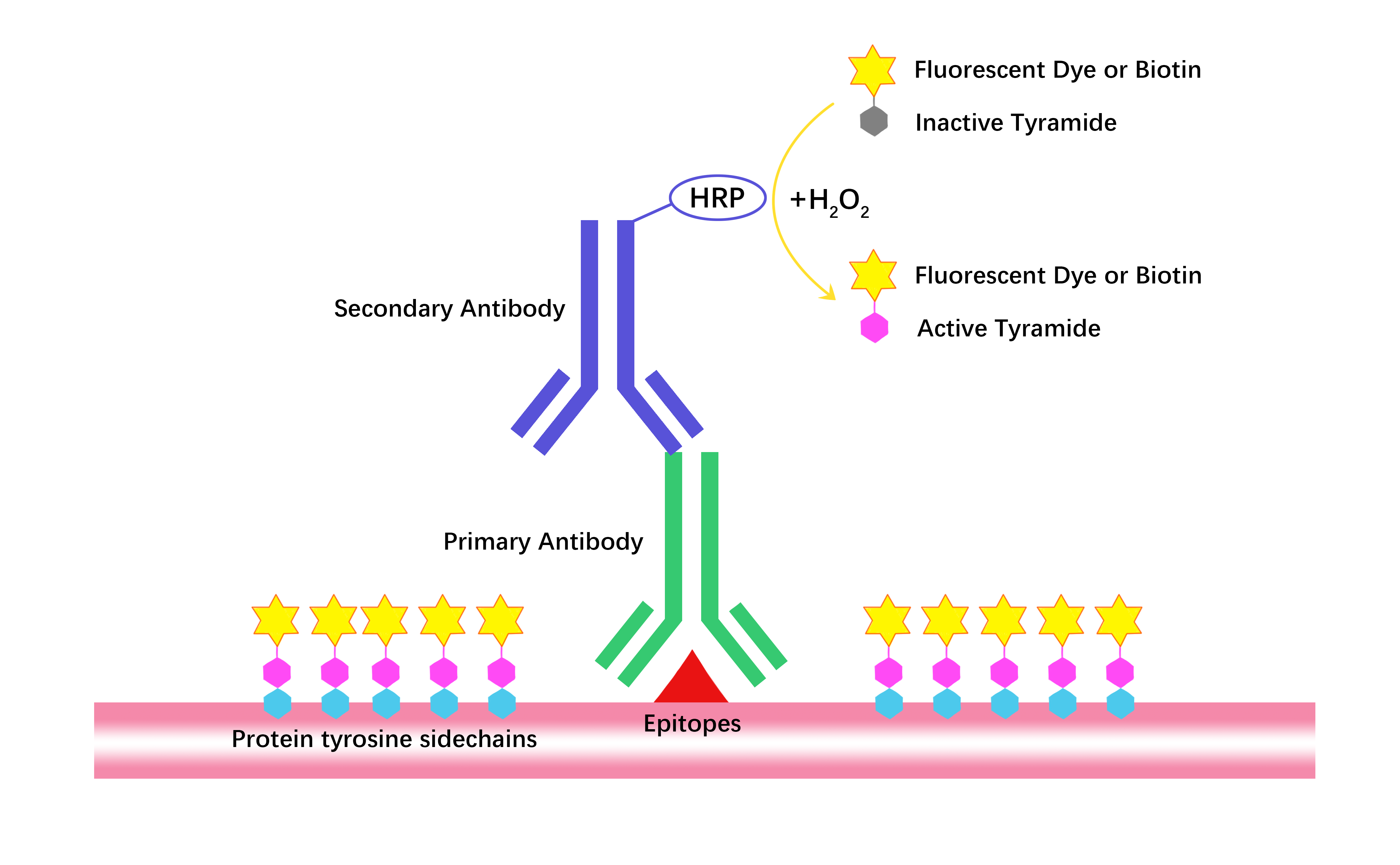


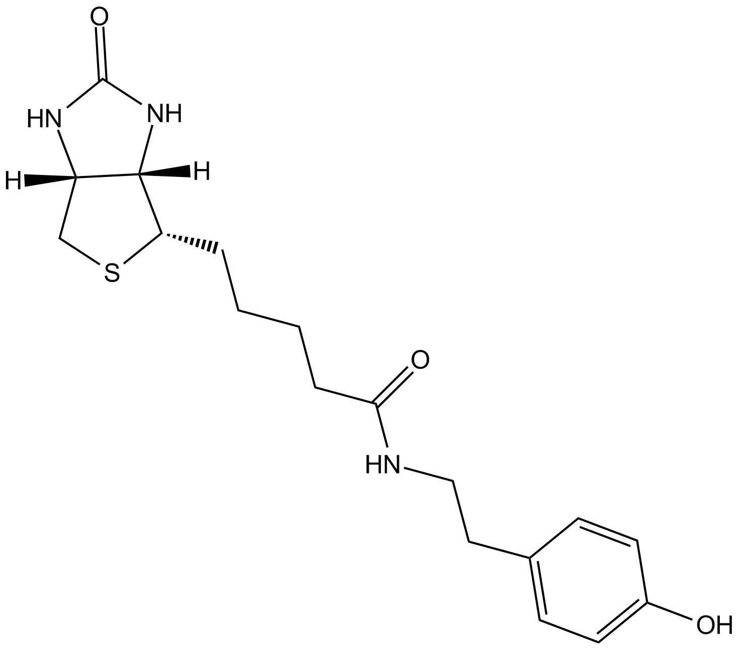
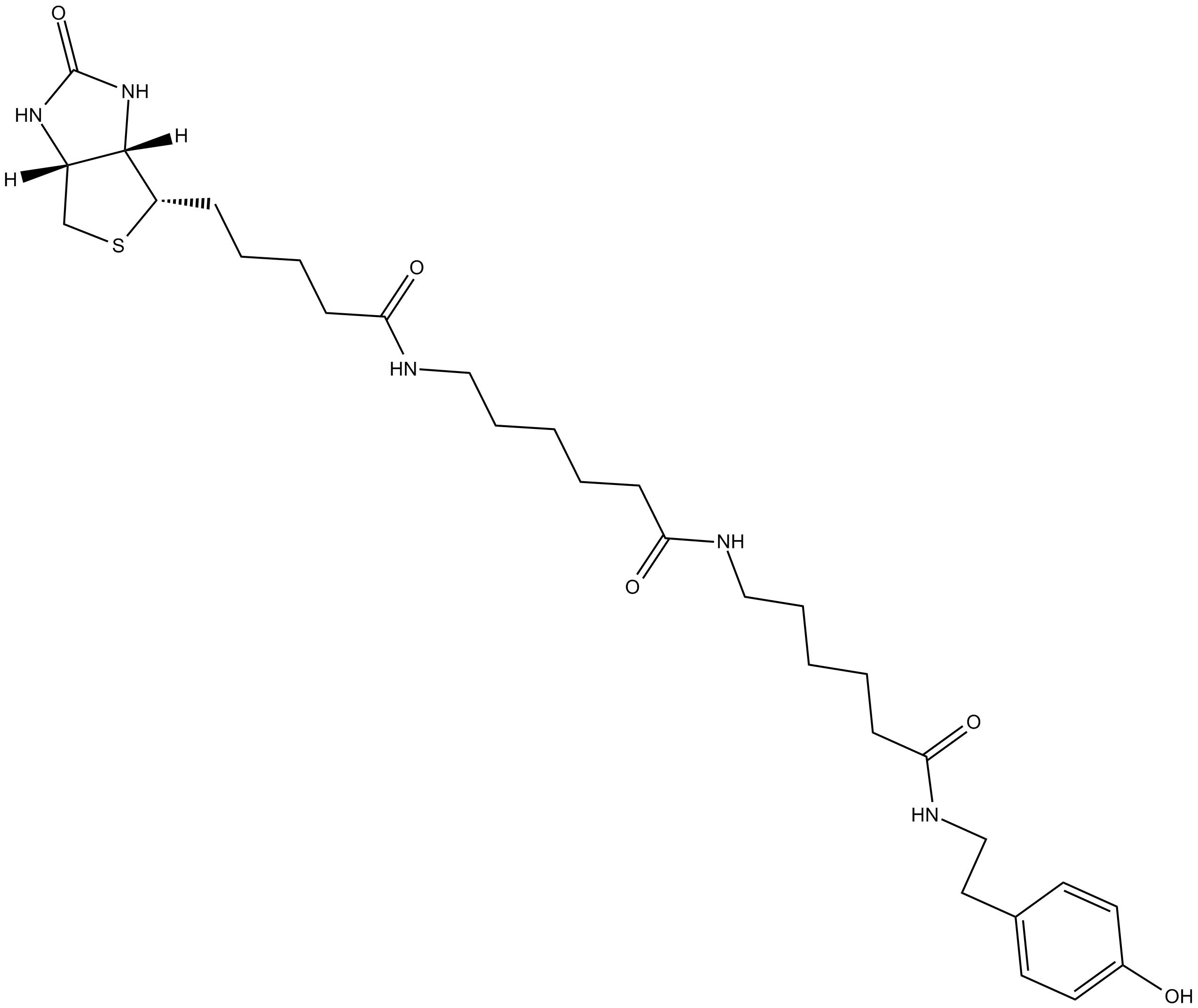
The Fluorescein TSA Fluorescence System uses tyramide signal amplification (TSA) to boost detection sensitivity in IHC, ICC, and ISH. HRP-linked secondary antibodies catalyze fluorescein-labeled tyramide into a reactive intermediate that covalently binds adjacent tyrosine residues, depositing high-density fluorescence around targets to amplify low-abundance molecule detection. Detectable via fluorescence microscopy (excitation 494 nm, emission 517 nm), it applies to analyzing proteins, nucleic acids, and other biomolecules in fixed cells and tissues.

- 1. Bo Xu, Xuezhou Ke, et al. "DNA From Neutrophil Extracellular Traps Restricts Group 3 Innate Lymphoid Cells Function in Intestinal Epithelial Repair via CCDC25." FASEB J. 2026 Jun 30;40(12):e72046. PMID: 42299911
- 2. Wang Jun, Chen Xiaoyang, et al. "Leonurine Ameliorates Doxorubicin-Induced Cardiotoxicity via STING/NF-κB/NLRP3 Inflammasome Signaling Pathway." Adv Sci (Weinh). 2026 Jun 12:e75912. PMID: 42283225
- 3. Alba Pau-Navalón, Tamara González-Costa, et al. "Endothelial USP8 is essential for angiogenesis." Angiogenesis. 2026 Jan 28;29(2):15. PMID: 41606310
- 4. Qiushi Feng, Xiaofeng Shan, et al. "Precision omic portrait deciphers the epigenetic variable during cellular identity reshaping of metastatic head and neck squamous cell carcinoma." Science Bulletin Available online 30 June 2026
- 5. Margaret E. Schroeder, Dana M. McCormack, et al. "A transcriptomic atlas of astrocyte heterogeneity across space and time in mouse and marmoset." Neuron. 2025 Nov 20:S0896-6273(25)00695-6. PMID: 41270736
- 6. Yiheng Mao, Yuan Li, et al. "All - at - once spatial proteome profiling of complex tissue context with single - cell - type resolution by proximity proteomics." Cell Syst. 2025 Jun 18;16(6):101291. PMID: 40345200
- 7. Chen Xiaoyang, Chen Yijun, et al. "Resibufogenin protects against atherosclerosis in ApoE-/-mice through blocking NLRP3 inflammasome assembly." J Adv Res. 2025 Apr 19:S2090-1232(25)00272-3. PMID: 40258472
- 8. Shaoyu Zhang, Qunxia Gu, et al. "An amino acid‐tuned gustatory receptor relatively abundant in the silkworm gut is crucial for growth and development." Pest Manag Sci. 2025 Apr 3. PMID: 40181590
- 9. Ke-Han Chen, Rui Xu, et al. "Evaluating the Efficacy and Safety of Emodin, Luteolin, and Paeonol Combination from Dahuang Mudan Decoction in Ameliorating Ulcerative Colitis." J Ethnopharmacol. 2025 Apr 25:346:119692. PMID: 40157404
- 10. Zengfeng Pan, Caiyan Gan, et al. "Gancao **exin decoction attenuated experimental colitis through suppressing ACSL4-mediated ferroptosis." J Ethnopharmacol. 2025 Mar 26:344:119532. PMID: 39993549
- 11. Xiaodong Duan, Chong Zhang, et al. "Suppression of epileptic seizures by transcranial activation of K+-selective channelrhodopsin." Nat Commun. 2025 Jan 10;16:559. PMID: 39789018
- 12. Qijun Wan, Zhichen Yang, et al. "Central Angiotensin II type 1 receptor deficiency alleviates renal fibrosis by reducing sympathetic nerve discharge in nephrotoxic folic acid–induced chronic kidney disease." PeerJ. 2024 Sep 26:12:e18166. PMID: 39346076
- 13. Xiaoxue Jiang, Kan liu, et al. "Hypothalamic SLC7A14 accounts for aging-reduced lipolysis in white adipose tissue of male mice." Nat Commun. 2024; 15: 7948. PMID: 39261456
- 14. Longyu Xu, Ruonan Ning, et al. "Bone Morphogenetic Protein Signaling Agonist SB4 (BMPSB4) Inhibits Corticotroph Pituitary Neuroendocrine Tumors by Activation of Autophagy via a BMP4/SMADs." ACS Pharmacol Transl Sci. 2024 Jun 24;7(7):1951-1970. PMID: 39022361
- 15. Jie Hong, Jie Liu, et al. "MiR-3180 inhibits hepatocellular carcinoma growth and metastasis by targeting lipid synthesis and uptake." Cancer Cell Int. 2023 Apr 11;23(1):66. PMID: 37041584
- 16. Jianan Li, Ruotian Xie, et al. "Tumor necrosis factor ligand‐related molecule 1A maintains blood–retinal barrier via modulating SHP‐1‐Src‐VE‐cadherin signaling in diabetic retinopathy." FASEB J. 2021 Nov;35(11):e22008. PMID: 34679191
Related Biological Data


| Complete Kit | 100-300 slides | 200-600 slides |
| 1X Amplification Diluent | 30 mL | 60 mL |
| Fluorescein Tyramide (dry, dissolve in 60 μL DMSO) | 1 tube | 2 tubes |
| Blocking Reagent | 6 g | 12 g |
Store Fluorescein Tyramide in the dark at -20°C for 2 years. Keep 1X Amplification Diluent and Blocking Reagent at 4°C for 2 years. | ||