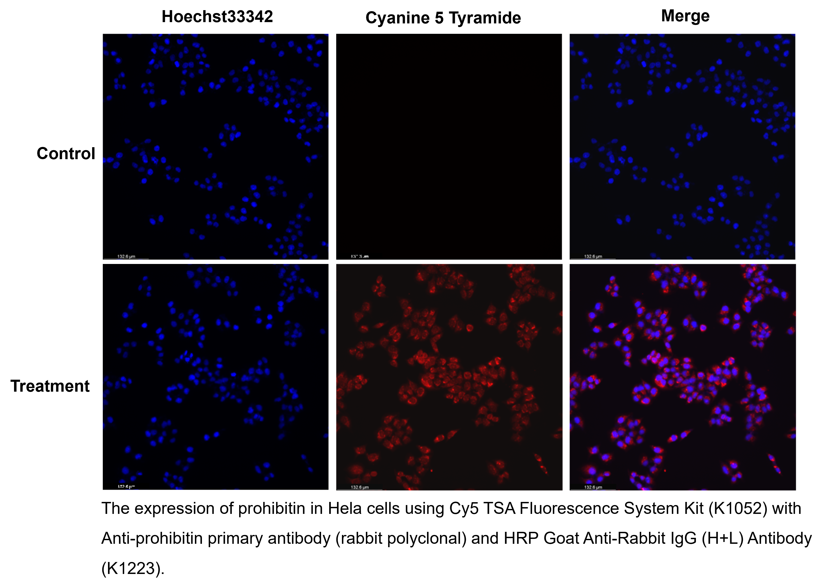
Cy5 TSA Fluorescence System Kit


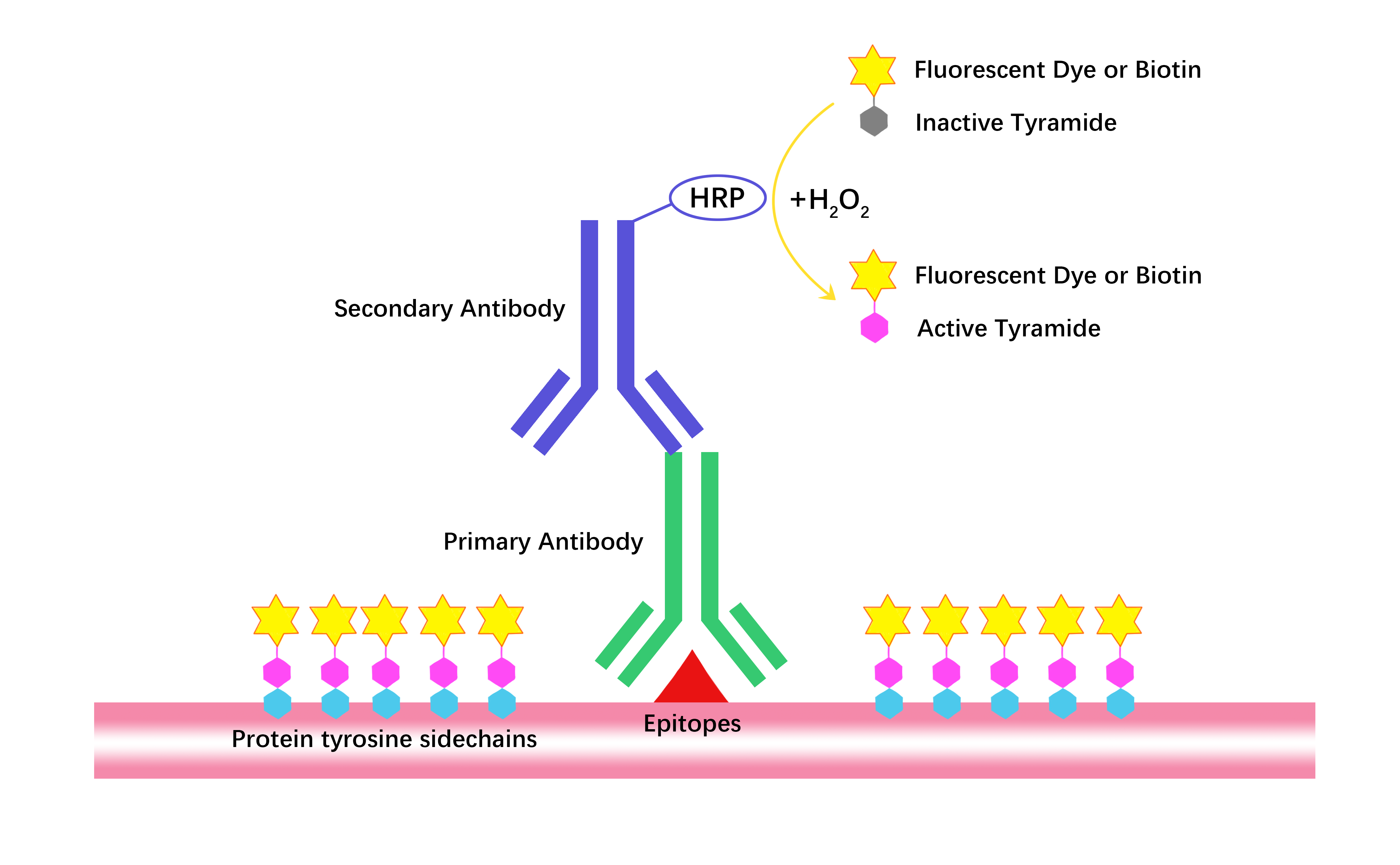


Tyramide Signal Amplification (TSA) from APExBIO technology increases sensitivity by a factor of 100 and enables detection of low-abundance targets in fluorescent immunocytochemistry (ICC), immunohistochemistry (IHC) and in situ hybridization (FISH) applications.
The TSA Fluorescence Kit uses horseradish peroxidase (HRP) to directly catalyze the covalent deposition of the fluorophore adjacent to the immobilized enzyme. The labeling process is so fast (less than ten minutes) and the deposition label can be viewed directly under standard or confocal microscopy. TSA fluorescein can also be used in combination with anti-fluorescein.
TSA technology is used for bright field microscopy of enzyme conjugates and suitable chromogenic substrates. The use of TSA reagents results in a significant increase in sensitivity compared to standard assays while maintaining stable specificity and resolution. In addition, TSA reagents can significantly reduce the consumption of primary antibodies or probes. The fluorescent labeling signal of this kit (K1052) is Cyanine 5, which can be detected the signal by microscopy at the excitation and emission wavelengths (648nm/667nm).
- 1. Margaret E. Schroeder, Dana M. McCormack, et al. "A transcriptomic atlas of astrocyte heterogeneity across space and time in mouse and marmoset." Neuron. 2025 Nov 20:S0896-6273(25)00695-6. PMID: 41270736
- 2. Jinsong Wei, Zhifa Cao, et al. "Nuclear ubiquitination permits Hippo–YAP signal for liver development and tumorigenesis." Nat Chem Biol. 2025 May 16. PMID: 40379800
- 3. Yiheng Mao, Yuan Li, et al. "All - at - once spatial proteome profiling of complex tissue context with single - cell - type resolution by proximity proteomics." Cell Syst. 2025 Jun 18;16(6):101291. PMID: 40345200
- 4. Chen Xiaoyang, Chen Yijun, et al. "Resibufogenin protects against atherosclerosis in ApoE-/-mice through blocking NLRP3 inflammasome assembly." J Adv Res. 2025 Apr 19:S2090-1232(25)00272-3. PMID: 40258472
- 5. Shaoting Jia, Weikang Lan, et al. "Functions of the transcription factor myb-like in sex determination and differentiation in Portunus trituberculatus." Aquaculture Volume 599, 15 April 2025, 742173
- 6. Yebin Wang, Zhenxing Zhong, et al. "Spatiotemporally restricted Hippo signalings instruct the fate and maturation of hepatobiliary cells." bioRxiv. November 03, 2024
- 7. Fanchu Zeng, Zhijin Fan, et al. "Tumor Microenvironment Activated Photoacoustic-Fluorescence Bimodal Nanoprobe for Precise Chemo-immunotherapy and Immune Response Tracing of Glioblastoma." ACS Nano. 2023 Oct 24;17(20):19753-19766. PMID: 37812513
- 8. Jie Hong, Jie Liu, et al. "MiR-3180 inhibits hepatocellular carcinoma growth and metastasis by targeting lipid synthesis and uptake." Cancer Cell Int. 2023 Apr 11;23(1):66. PMID: 37041584
- 9. Horwitz, Lorraine. "Novel Sensory Pathways for Thermosensation and Pain Under Physiological and Pathological Conditions." University of Michigan Library. 2022
- 10. Huayue Li, Zhongwu Chen, et al. "MiR-4310 regulates hepatocellular carcinoma growth and metastasis through lipid synthesis." Cancer Lett. 2021 Oct 28;519:161-171. PMID: 34303763
Quality Control & DataSheet
- View current batch:
-
Purity = 99.45%
- COA (Certificate Of Analysis)
- HPLC
- Protocol
Related Biological Data

Related Biological Data

| Complete Kit | 100-300 slides | 200-600 slides |
| 1X Amplification Diluent | 30 mL | 60 mL |
| Cyanine 5 Tyramide (dry, dissolve in 60 μL DMSO) | 1 tube | 2 tubes |
| Blocking Reagent | 6 g | 12 g |
Store Cyanine 5 Tyramide in the dark at -20°C for 2 years. Keep 1X Amplification Diluent and Blocking Reagent at 4°C for 2 years. | ||