Nile Red


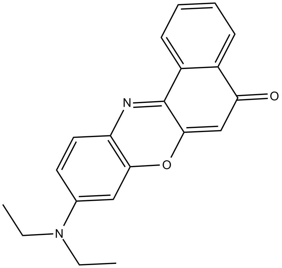
Nile Red is a lipophilic fluorescent compound, also known as Nile blue oxazone, with CAS number 7385-67-3 and a molecular weight of 318.37. Its maximum excitation wavelength in phospholipids is approximately 552nm, with a maximum emission wavelength of about 636nm, producing red fluorescence; it can also be excited at wavelengths of 450-500nm with emission set to greater than 528nm, resulting in green fluorescence. However, red fluorescence is usually much brighter than green fluorescence. Since Nile Red can stain both cell membranes and lipid droplets during red fluorescence, but only stains lipid droplets during green fluorescence, if there is a particular focus on the specificity of lipid droplets, the parameters can be set to Ex/Em=485/535nm for detection. Using Nile Red, one can study lipid distribution, lipid metabolism, and lipid storage dynamics in different cell types, aiding in the analysis of lipid-related physiological and pathological processes.
- 1. Shaokun Huang, Xuan Shan, et al. "Integrative analysis identified THBS1 as a key prognostic biomarker with therapeutic vulnerability in patients with laryngeal cancer." Transl Cancer Res. 2025 Oct 31;14(10):6723-6737. PMID: 41234877
- 2. Penelope E. Jankoski, Abdul-Razak Masoud, et al. "Bioactive Supramolecular Polymers for Skin Regeneration Following Burn Injury." Biomacromolecules. 2025 Aug 11;26(8):5471-5482. PMID: 40669455
- 3. Penelope E. Jankoski, Zacchaeus M. Wallace, et al. "Combating Reactive Oxygen Species (ROS) with Antioxidant Supramolecular Polymers." ACS Appl Mater Interfaces. 2025 Jun 4;17(24):35275–35287. PMID: 40462549
- 4. Qiushi Zheng, Chao Li, et al. "Candida auris cells form giant lipid droplets to survive in harsh environments." Commun Biol. 2025 May 22;8(1):783. PMID: 40404799
- 5. Ronit Vogt Sionov, Maya Korem, et al. "Cannabidiol (CBD) Acts as an Antioxidant on Gardnerella vaginalis, Resulting in Reduced Metabolic Activity, Loss of Survivability, and Elimination of Biofilms." Antibiotics (Basel). 2025 Feb 1;14(2):136. PMID: 40001381
- 6. Ronit Vogt Sionov, et al. "Anti-Bacterial and Anti-Biofilm Activities of Arachidonic Acid against the Cariogenic Bacterium Streptococcus mutans." Front Microbiol. 2024 Feb 26:15:1333274. PMID: 38596377
- 7. Ye Zhu, et al. "Metabarcoding Analysis of Microorganisms Inside Household Washing Machines in Shanghai, China." Microorganisms. 2024 Jan 13;12(1):160. PMID: 38257987
- 8. Jun Zhao, Rui Wang, et al. "Suppression of AQP7 is Crucial for Proliferation and Lipid Metabolism in ccRCC." Research Square. 13 Mar, 2024
- 9. Nathanyel Sebbane, Itzhak Abramovitz, et al. "Mechanistic Insight into the Anti-Bacterial/Anti-Biofilm Effects of Low Chlorhexidine Concentrations on Enterococcus faecalis—In Vitro Study." Microorganisms 2024, 12(11), 2297
- 10. Athira Venugopal, Ronit Vogt Sionov, et al. "The V-Shaped Structuring Regulated via the LuxS-Dependent Quorum-Sensing Pathway Is Associated With Lactiplantibacillus plantarum Survivability in Acidic Environments." FOOD FRONTIERS. 25 November 2024
- 11. Badri Parshad, Andrew George Baker, et al. "Improved Therapeutic Efficiency of Senescent Cell-specific, Galactose-Functionalized Micelle Nanocarriers." SMALL. 18 December 2024
- 12. Goldie Wolfson, Ronit Vogt Sionov, et al. "Anti-Bacterial and Anti-Biofilm Activities of Anandamide against the Cariogenic Streptococcus mutans." Int J Mol Sci. 2023 Mar 24;24(7):6177. PMID: 37047147
| Storage | Store at -20°C |
| M.Wt | 318.37 |
| Cas No. | 7385-67-3 |
| Formula | C20H18N2O2 |
| Solubility | ≥2.56 mg/mL in DMSO; insoluble in EtOH; insoluble in H2O |
| Chemical Name | 9-(diethylamino)-5H-benzo[a]phenoxazin-5-one |
| Canonical SMILES | CCN(CC)c(cc1O2)ccc1N=C(c1c3cccc1)C2=CC3=O |
| Shipping Condition | Small Molecules with Blue Ice, Modified Nucleotides with Dry Ice. |
| General tips | We do not recommend long-term storage for the solution, please use it up soon. |
| Cell experiment:[1] | |
|
Cell lines |
Monkey aortic smooth muscle cells and mouse peritoneal macrophages, induced by acetylated low density lipoprotein |
|
Reaction Conditions |
100 ng/ml Nile Red |
|
Applications |
Better selectivity for cytoplasmic lipid droplets was obtained when the cells were viewed for yellow-gold fluorescence (excitation, 450-500 nm; emission, greater than 528 nm) rather than red fluorescence (excitation, 515-560 nm; emission, greater than 590 nm). |
|
Note |
The technical data provided above is for reference only. |
|
References: 1. Greenspan P, Mayer EP, Fowler SD. Nile red: a selective fluorescent stain for intracellular lipid droplets. Journal of Cell Biology, 1985, 100(3): 965-973. |
|
Quality Control & MSDS
- View current batch:
Chemical structure