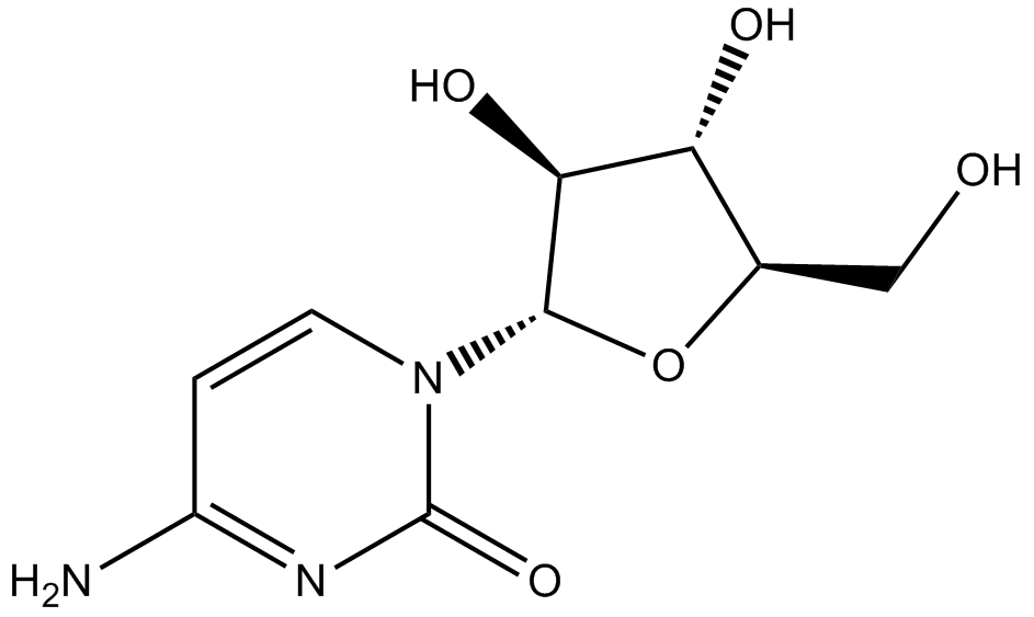
Cytarabine


Cytarabine (CAS 147-94-4), also known as AraC, is a nucleoside analog structurally related to deoxycytidine. It acts primarily by incorporation into DNA, subsequently hindering DNA synthesis via inhibition of DNA and RNA polymerases. AraC requires phosphorylation by deoxycytidine kinase (dCK) to its active monophosphate metabolite. Reduced dCK activity or expression of alternatively spliced inactive dCK isoforms has been linked to resistance in leukemic cell lines. Additionally, cytarabine induces apoptosis and inhibits cell proliferation, involving p53 stabilization independent of transcriptional elevation as demonstrated in rat trophoblast cells. Clinically, cytarabine remains extensively studied in leukemia models and cellular apoptosis pathways.
- 1. Yang Li, Hanying Huang, et al. "TSC22D3 as an immune-related prognostic biomarker for acute myeloid leukemia." iScience. 2023 Jul 22;26(8):107451. PMID: 37575189
- 2. Xiahong Tang, Jun Ke, et al. "Hypoxic preconditioned mesenchymal stem cells ameliorate rat brain injury after cardiopulmonary resuscitation by suppressing neuronal pyroptosis." J Cell Mol Med. 2023 Jul;27(13):1836-1858. PMID: 37246833
- 3. Jing-Ting Chiou, Chia-Chi Hsu, et al. "Cytarabine-induced destabilization of MCL1 mRNA and protein triggers apoptosis in leukemia cells." Biochem Pharmacol. 2023 May:211:115494. PMID: 36924905
- 4. Emmanuelle Supper, Saskia Rudat, et al. "Cut-like homeobox 1 (CUX1) tumor suppressor gene haploinsufficiency induces apoptosis evasion to sustain myeloid leukemia." Nat Commun. 2021 Apr 30;12(1):2482. PMID: 33931647
- 5. Zhijun Liu, Himani Nailwal, et al. "A class of viral inducer of degradation of the necroptosis adaptor RIPK3 regulates virus-induced inflammation." Immunity. 2021 Feb 9;54(2):247-258.e7. PMID: 33444549
- 6. Lin KH, Xie A, et al. "Systematic Dissection of the Metabolic-Apoptotic Interface in AML Reveals Heme Biosynthesis to Be a Regulator of Drug Sensitivity." Cell Metab. 2019 Feb 5. pii: S1550-4131(19)30011-7. PMID: 30773463
| Physical Appearance | A solid |
| Storage | Store at -20°C |
| M.Wt | 243.2 |
| Cas No. | 147-94-4 |
| Formula | C9H13N3O5 |
| Solubility | insoluble in EtOH; ≥28.6 mg/mL in H2O; ≥11.73 mg/mL in DMSO |
| Chemical Name | 4-amino-1-[(2R,3S,4S,5R)-3,4-dihydroxy-5-(hydroxymethyl)oxolan-2-yl]pyrimidin-2-one |
| Canonical SMILES | O=C1N=C(C=CN1[C@H]2[C@@H](O)[C@H](O)[C@@H](CO)O2)N |
| Shipping Condition | Small Molecules with Blue Ice, Modified Nucleotides with Dry Ice. |
| General tips | We do not recommend long-term storage for the solution, please use it up soon. |
| Cell experiment [1]: | |
|
Cell lines |
rat sympathetic neurons |
|
Preparation method |
Limited solubility in DMSO. General tips for obtaining a higher concentration: Please warm the tube at 37 ℃ for 10 minutes and/or shake it in the ultrasonic bath for a while. Stock solution can be stored below -20℃ for several months. |
|
Reacting condition |
10 μM |
|
Applications |
Cytarabine apparently induced apoptosis of rat sympathetic neurons at 10 μM, of which 100 μM showed the highest toxicity and killed over 80% of the neurons by 84 hours, involving the release of mitochondrial cytochrome-c and the activation of caspase-3. |
| Animal experiment [2]: | |
|
Animal models |
Pregnant Slc:Wistar rats |
|
Dosage form |
Intraperitoneal injection, 250 mg/kg |
|
Application |
Cytarabine (250 mg/kg) caused placental growth retardation and increased placental trophoblastic cells apoptosis in the placental labyrinth zone of the pregnant Slc:Wistar rats, which increases from 3 hour after the treatment and peaks at 6 hour before returning to control levels at 48 hour, with remarkably enhanced p53 protein, p53 trancriptional target genes such as p21, cyclinG1 and fas and caspase-3 activity. |
|
Other notes |
Please test the solubility of all compounds indoor, and the actual solubility may slightly differ with the theoretical value. This is caused by an experimental system error and it is normal. |
|
References: [1]. Besirli C G, Deckwerth T L, Crowder R J, et al. Cytosine arabinoside rapidly activates Bax-dependent apoptosis and a delayed Bax-independent death pathway in sympathetic neurons[J]. Cell death and differentiation, 2003, 10(9): 1045. [2]. Yamauchi H, Katayama K, Ueno M, et al. Involvement of p53 in 1-β-D-arabinofuranosylcytosine-induced trophoblastic cell apoptosis and impaired proliferation in rat placenta[J]. Biology of reproduction, 2004, 70(6): 1762-1767. |
|
Quality Control & MSDS
- View current batch:
Chemical structure
Related Biological Data