Recombinant Human M-CSF/CSF1
Macrophage Colony Stimulating Factor(M-CSF), also known as CSF-1, is a four-alpha -helical-bundle cytokine that is the primary regulator of macrophage survival, proliferation and differentiation [1-3]. M-CSF is also essential for the survival and proliferation of osteoclast progenitors [1, 4]. M-CSF also primes and enhances macrophage killing of tumor cells and microorganisms, regulates the release of cytokines and other inflammatory modulators from macrophages, and stimulates pinocytosis [2, 3]. M-CSF increases during pregnancy to support implantation and growth of the decidua and placenta [5]. Sources of M-CSF include fibroblasts, activated macrophages, endometrial secretory epithelium, bone marrow stromal cells and activated endothelial cells [1-5]. The M-CSF receptor (c-fms) transduces its pleotropic effects and mediates its endocytosis. M-CSF mRNAs of various sizes occur [3-9]. Full length human M-CSF transcripts encode a 522 amino acid (aa) type I transmembrane (TM) protein with a 464 aa extracellular region, a 21 aa TM domain, and a 37 aa cytoplasmic tail that forms a 140 kDa covalent dimer. Differential processing produces two proteolytically cleaved, secreted dimers. One is an N- and O- glycosylated 86 kDa dimer, while the other is modified by both glycosylation and chondroitin-sulfate proteoglycan (PG) to generate a 200 kDa subunit. Although PG-modified M-CSF can circulate, it may be immobilized by attachment to type V collagen [8]. Shorter transcripts encode M-CSF that lacks cleavage and PG sites and produces an N-glycosylated 68 kDa TM dimer and a slowly produced 44 kDa secreted dimer [7]. Although forms may vary in activity and half-life, all contain the N-terminal 150 aa portion [10].
Reference
[1]. Pixley, F.J. and E.R. Stanley (2004) Trends Cell Biol. 14:628.
[2]. Chitu, V. and E.R. Stanley (2006) Curr. Opin. Immunol. 18:39.
[3]. Fixe, P. and V. Praloran (1997) Eur. Cytokine Netw. 8:125.
[4]. Ryan, G.R. et al. (2001) Blood 98:74.
[5]. Makrigiannakis, A. et al. (2006) Trends Endocrinol. Metab. 17:178.
[6]. Nandi, S. et al. (2006) Blood 107:786.
[7]. Rettenmier, C.W. and M.F. Roussel (1988) Mol. Cell Biol. 8:5026.
[8]. Suzu, S. et al. (1992) J. Biol. Chem. 267:16812.
[9]. Manos, M.M. (1988) Mol. Cell. Biol. 8:5035.
[10]. Koths, K. (1997) Mol. Reprod. Dev. 46:31.
|
Accession # |
P09603 |
|
Alternate Names |
Colony stimulating factor 1 (macrophage); CSF1; CSF-1; macrophage colony-stimulating factor 1; MCSF |
|
Source |
Human embryonic kidney cell, HEK293-derived human M-CSF/CSF1 protein |
|
Protein sequence |
Glu33-Ser190 |
|
M.Wt |
22.0 kDa(Monomer) |
|
Appearance |
Solution protein. |
|
Stability & Storage |
Avoid repeated freeze-thaw cycles. It is recommended that the protein be aliquoted for optimal storage. 3 years from date of receipt, -20 to -70 °C as supplied. |
|
Concentration |
0. 2 mg/mL |
|
Formulation |
Dissolved in sterile PBS buffer. |
|
Reconstitution |
We recommend that this vial be briefly centrifuged prior to opening to bring the contents to the bottom. This solution can be diluted into other aqueous buffers. |
|
Biological Activity |
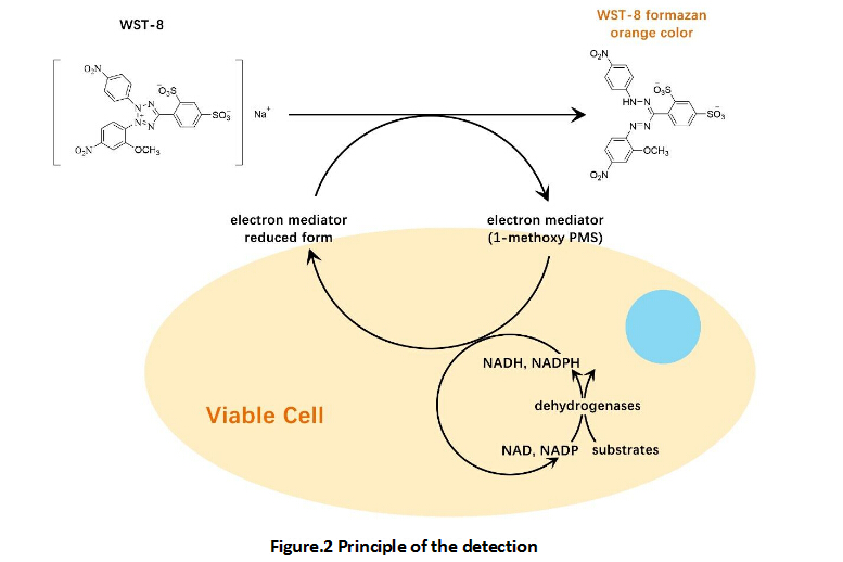
Measured in a cell proliferation assay using M-NFS-60 mouse myelogenous leukemia lymphoblast cells. The EC50 for this effect is 0.2-1.5 ng/mL. |
|
Shipping Condition |
Shipping with dry ice. |
|
Handling |
Centrifuge the vial prior to opening. |
|
Usage |
For Research Use Only! Not to be used in humans. |
Quality Control & DataSheet
- View current batch:
-
Purity > 95%, determined by SDS-PAGE.
- Datasheet
Endotoxin: <0.010 EU per 1 ug of the protein by the LAL method.


