CFDA-SE


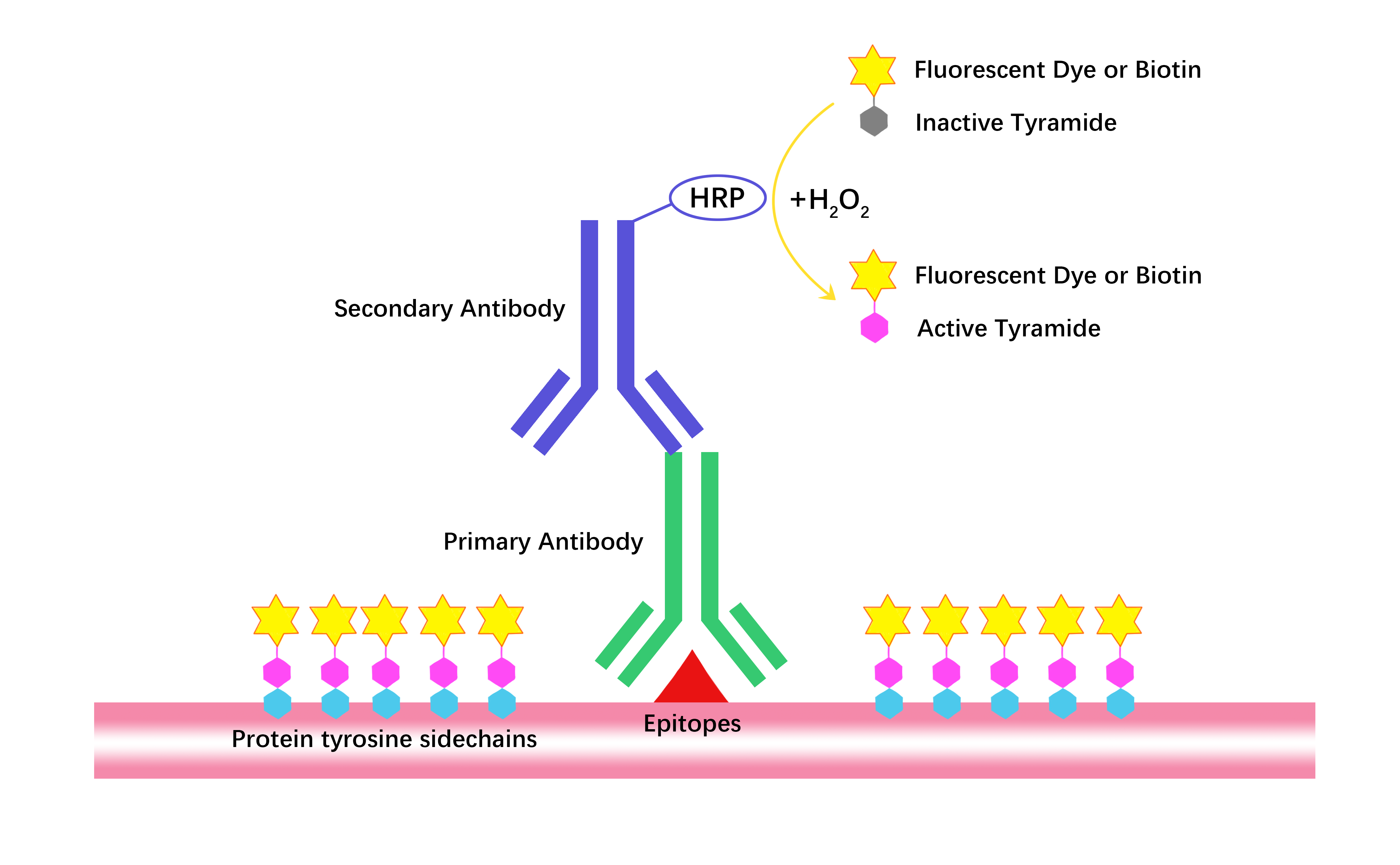


CFDA-SE (Carboxyfluorescein diacetate succinimidyl ester; CAS: 150347-59-4) is a membrane-permeant fluorescent dye commonly utilized for labeling viable cells and monitoring cellular proliferation. Upon entering cells, intracellular esterases hydrolyze CFDA-SE into carboxyfluorescein succinimidyl ester (CFSE), which becomes fluorescent (excitation 494 nm, emission 521 nm), binds covalently to intracellular amino groups, and is thereafter trapped within the cell cytoplasm. Each cell division results in halving fluorescence intensity, enabling accurate quantification of successive proliferation events via flow cytometry. CFDA-SE labeling methodology has extensive applications in assessing lymphocyte, fibroblast, NK cell, and bacterial proliferation both in vitro and in vivo.
| Storage | -20℃, sealed storage, away from moisture and light |
| M.Wt | 557.46 |
| Cas No. | 150347-59-4 |
| Formula | C29H19NO11 |
| Solubility | ≥37.17 mg/mL in DMSO with ultrasonic; insoluble in EtOH; insoluble in H2O |
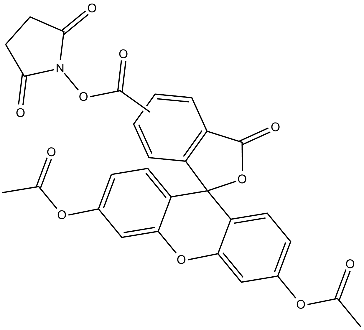
| Chemical Name | 3',6'-bis(acetyloxy)-3-oxo-2,5-dioxo-1-pyrrolidinyl ester-spiro[isobenzofuran-1(3H),9'-[9H]xanthene]-ar-carboxylic acid |
| Canonical SMILES | CC(OC1=CC2=C(C3(C4=C(C=C(OC(C)=O)C=C4)O2)C5=C(C(O3)=O)C=C(C(ON6C(CCC6=O)=O)=O)C=C5)C=C1)=O.CC(OC7=CC8=C(C9(C%10=C(C(O9)=O)C=CC(C(ON%11C(CCC%11=O)=O)=O)=C%10)C%12=C(C=C(OC(C)=O)C=C%12)O8)C=C7)=O |
| Shipping Condition | Small Molecules with Blue Ice, Modified Nucleotides with Dry Ice. |
| General tips | We do not recommend long-term storage for the solution, please use it up soon. |
| Cell experiment:[1] | |
|
Cell lines |
Human erythroleukaemic cell line K562, mouse lymphoma cell line YAC-1, human mammary cancer cell line MCF-7 and human melanoma cell line A375 |
|
Reaction Conditions |
1, 1.5, 2, 2.5, 5 and 10 µM CFDA-SE |
|
Applications |
CFDA-SE at 2.5 μM stained more than 95% of the cells on all cell lines tested, and dose-dependently increased fluorescence intensity of stained cells. The optimal concentration for K562 and YAC-1 was found to be 2.5 μM, while the optimal concentrations for A375 and MCF-7 were found to be 5 μM and 10 μM, respectively. Within the 6 h experiment period, no cytotoxicity related to CFDA-SE was observed. |
| Animal experiment:[4] | |
|
Animal models |
C57BL/6 mice, aged 5 ~ 8 weeks |
|
Dosage form |
10 μM Injected into thymic lobe |
|
Applications |
CFDA-SE, at 80 times the concentration used for in vitro labeling, was nontoxic and labeled randomly approximately 15% of thymocytes 24 h after injection. The turnover rate of labeled thymic emigrants in the lymph nodes was in the order of 21 days. Thus, CFDA-SE may serve as a powerful tool in relatively long-term migration studies. |
|
Note |
The technical data provided above is for reference only. |
|
References: 1. Wang XQ, Duan XM, Liu LH, et al. Carboxyfluorescein diacetate succinimidyl ester fluorescent dye for cell labeling. Acta Biochimica et Biophysica Sinica (Shanghai), 2005, 37(6): 379-385. 2. Weston SA, Parish CR. New fluorescent dyes for lymphocyte migration studies. Analysis by flow cytometry and fluorescence microscopy. Journal of Immunological Methods, 1990, 133(1): 87-97. 3. Parish CR, Glidden MH, Quah BJ, et al. Use of the intracellular fluorescent dye CFSE to monitor lymphocyte migration and proliferation. Current Protocols in Immunology, 2009, Chapter 4: Unit4.9. 4. Graziano M, St-Pierre Y, Beauchemin C, et al. The fate of thymocytes labeled in vivo with CFSE. Experimental Cell Research, 1998, 240(1): 75-85. |
|
Quality Control & MSDS
- View current batch:
Chemical structure