ECL Chemiluminescent Substrate Detection Kit (Hypersensitive)

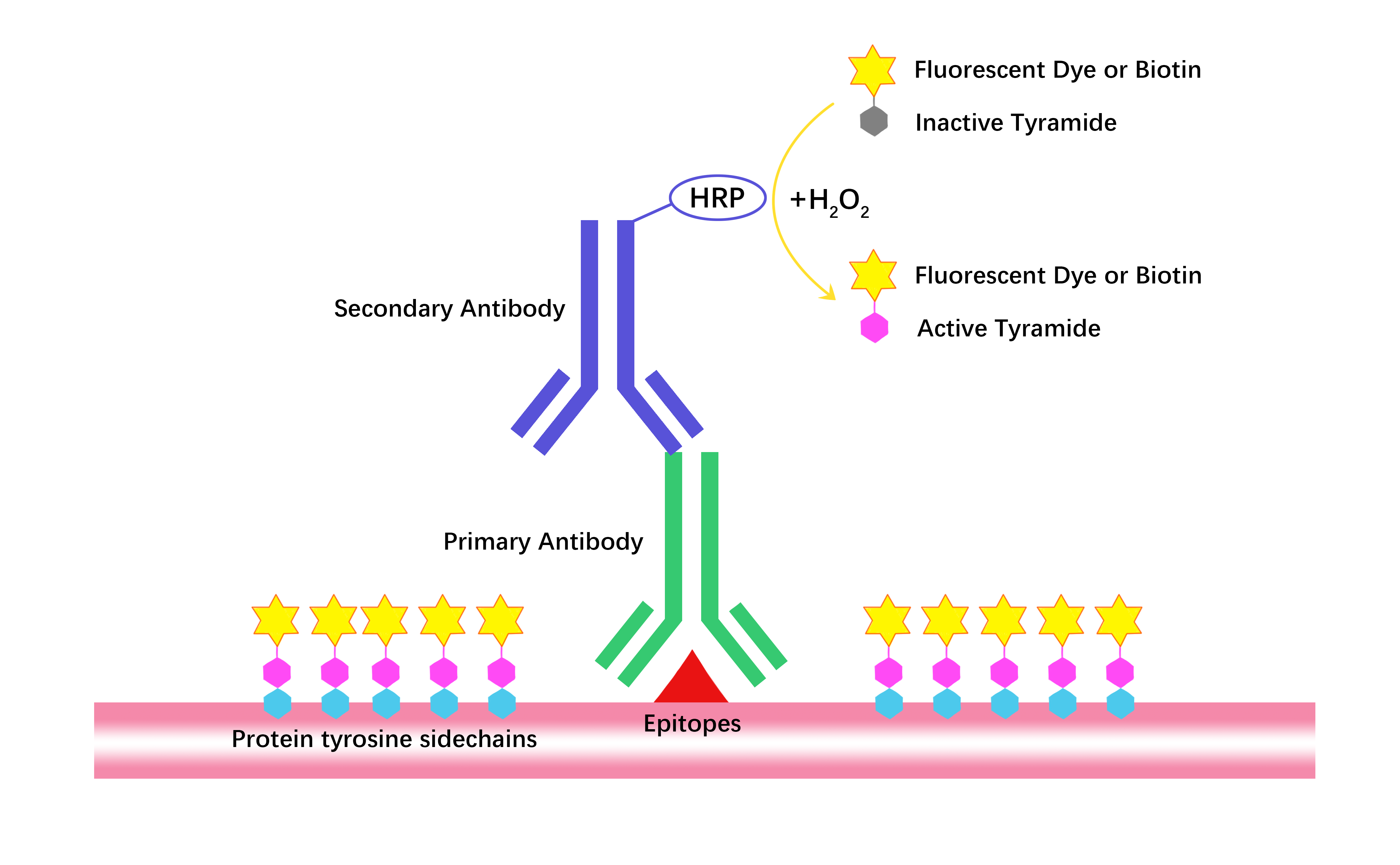


ECL Chemiluminescent Substrate Detection Kit (Hypersensitive), using HRP-mediated oxidation to generate chemiluminescent signals, is for immunoblotting to detect antigens on nitrocellulose/PVDF membranes, with high sensitivity (low picogram range) for low-abundance proteins. Its signals persist for 6-8 hours under optimal conditions, and the mixed working reagent is stable for 24 hours. It has an extended room-temperature shelf-life, and compared to conventional kits (e.g., from T and M), it shows lower background and longer signal duration.
Product Features
(1) Enhanced chemiluminescent substrate for horseradish peroxidase (HRP).
(2) Low picogram sensitivity - Detects low picogram protein bands on nitrocellulose or PVDF membranes.
(3) Long signal duration - Under optimized conditions, substrate-incubated blots can consistently output a detectable light signal for 6 to 8 hours.
(4) Stable reagent - The working solution remains stable for 24 hours; The kit can be stored at room temperature for up to 1 year.
(5) Cost-effective - Optimized for diluted antibody concentrations.
- 1. Wenzhen Gao, Li Zhu, et al. "BMAL1 regulates circadian rhythms via phase separation–mediated transcriptional hub formation." Signal Transduct Target Ther. 2026 May 1;11(1):160. PMID: 42062246
- 2. Wen-Bo Cui, Xin-Yu Meng, et al. "Broussonetia papyrifera Fruit Extract Attenuates Alzheimer's Pathogenesis via Disrupting the Vicious Cycle of Aβ and Oxidative Stress in C. elegans and Cellular models." Journal of Ethnopharmacology Volume 364, 12 June 2026, 121513. PMID: 41819509
- 3. Shuang Lu, Lewen Wang, et al. "Acetylation of PRDX5 aggravates the oxidative stress and apoptosis of retinal neurons induced by ischemia-reperfusion." Tissue Cell. 2026 Feb 23:101:103408. PMID: 41740330
- 4. Long Cheng, Minghong Gan, et al. "Puerarin Improves Glucose and Lipid Metabolism in Type 2 Diabetes by Regulating Gut Microbiota Homeostasis and Promoting Adipose Tissue Thermogenesis." Phytother Res. 2026 Feb 16. PMID: 41696817
- 5. Chen Li, Yike Li, et al. "Unveiling the neuroprotective power of mitochondrial transfer in orofacial inflammatory pain through ER membrane remodeling." Cell Reports Volume 45, Issue 1, 27 January 2026, 116809. PMID: 41538328
- 6. Shuang Lu, Lewen Wang, et al. "As a novel ferroptosis inhibitor, primaquine alleviates I/R-induced retinal neuron death via increasing GSTA1 activity." Biomedicine & Pharmacotherapy Volume 195, February 2026, 118973. PMID: 41529513
- 7. Xingpeng Wang, Lian Zhong, et al. "Target Validation and Drug Discovery for TNBC: Targeting the Lin28B/Let-7/PBK with the Ponicidin." European Journal of Medicinal Chemistry Volume 304, 15 February 2026, 118474. PMID: 41406550
- 8. Hongyao Li, Xiaoqi Wu, et al. "Gastrodin Alleviates Tau Hyperphosphorylation Associated with AKT/GSK-3β Signaling Changes in an Alzheimer's Disease Cell Model." JoVE Journal March 24, 2026
- 9. Jingtian Mu, Tingpei Ye, Junjiang Liu. "CAFs-secreted Fatty Acids Fuel Oral Cancer Progression via Lipid Raft Formation." Arch Oral Biol . 2025 Aug 15:179:106377. PMID: 40840065
- 10. Qi Zhang, Ruiqi Wang, et al. "A humanized Gs-coupled DREADD for circuit and behavior modulation." Front Cell Neurosci. 2025 Apr 9;19:1577117. PMID: 40271540
- 11. Yan-Yun Liu, Zheng-Ao Li, et al. "TCM theory-inspired discovery of DNJ-flavonoid conjugates as broad-spectrum anti-SARS-CoV-2 agents by primarily targeting ER-associated glycoprotein folding process." Eur J Med Chem. 2025 Jun 5:290:117582. PMID: 40168909
- 12. Zhina Wu, Jianai Chen, et al. "Polypeptide of Inonotus hispidus extracts alleviates periodontitis through suppressing inflammatory bone loss." Int J Biol Macromol. 2025 Jan;287:138350. PMID: 39645101
- 13. Jie Hao, Yanfeng Zhu, et al. "Structural characterization and hypolipidemic activity of a hetero-galactan purified from Sanghuangporus vaninii based on modulation of TLR4/NF-κB pathway." Carbohydr Polym. 2025 Jan 1:347:122702. PMID: 39486943
- 14. ZHINA WU, RUI LIU, et al. "An enzymatic cleavage-triggered minimally invasive nanosensor for urine-based detection of early atherosclerosis." SCIENCE ADVANCES 14 Mar 2025 Vol 11, Issue 11
- 15. Jiajing Cai, Bei Xia, et al. "Site-Specific DNA methylation detection using nonlinear hybridization chain reaction." Microchemical Journal. Volume 212, May 2025, 113522
- 16. Weiyun Wu, Xiaowen Li, et al. "METTL14 regulates inflammation in ulcerative colitis via the lncRNA DHRS4-AS1/miR-206/A3AR axis." Cell Biol Toxicol 40, 95 (2024) PMID: 39528760
- 17. Weiyun Wu, Aiting Li, et al. "Long noncoding RNA LINC01550 inhibits colorectal cancer malignancy by suppressing the Wnt/β‐catenin signaling pathway." J Biochem Mol Toxicol. 2024 Aug;38(8):e23774. PMID: 39041324
Related Biological Data
")
Related Biological Data
")
Related Biological Data
")
Related Biological Data
")
| Components | 100 mL | 500 mL |
| ECL Chemiluminescent Substrate Detection Kit (Hypersensitive)-A | 50 mL | 250 mL |
| ECL Chemiluminescent Substrate Detection Kit (Hypersensitive)-B | 50 mL | 250 mL |
Store the components dry at 4 °C and protect from light for 12 months. | ||
1. What causes an inverted image on the film, i.e., black background with white bands?
A: This is usually caused by an excessive amount of HRP in the system, which rapidly depletes the substrate. It is recommended to dilute the HRP-conjugated secondary antibody at least 10-fold to effectively resolve this issue.
2. What causes brown or yellow bands on the membrane?
A: This phenomenon is often related to suboptimal chemiluminescent development conditions, such as overly long substrate incubation or exposure times, or substrate degradation/precipitation under light. Shortening the development/exposure time and optimizing the detection conditions can alleviate this issue.
3. If the chemiluminescent signal lasts less than 8 hours, what could be the reason?
A: An excessively short signal duration is usually caused by too much HRP in the system, which rapidly depletes the substrate and leads to quick signal decay. The solution is to dilute the HRP-conjugated secondary antibody at least 10-fold.
4. If the signal is weak or absent, what are the possible causes and how can this be resolved?
A: Common causes and corresponding solutions include:
· Excessive HRP in the system (substrate depleted quickly, signal decays rapidly): dilute the HRP-conjugated secondary antibody at least 10-fold.
· Insufficient amount of antigen or antibody: increase the concentration of antigen or antibody accordingly.
· Low protein transfer efficiency: optimize transfer conditions (e.g., transfer time, current/voltage, buffer composition).
· Reduced HRP or substrate activity: perform activity testing to confirm and replace inactivated reagents.
5. If the background is too high, what are the possible causes and how can this be resolved?
A: Common causes of high background and corresponding solutions include:
· Excessive HRP in the system: dilute the HRP-conjugated secondary antibody at least 10-fold.
· Insufficient blocking: optimize blocking conditions, such as extending blocking time, adjusting blocking temperature, or shaking speed.
· Inappropriate blocking reagent: try different blocking reagents, such as non-fat dry milk, BSA, or serum.
· Inadequate washing: increase the number of washes, extend washing time, or increase the volume of wash buffer.
· Overexposure of the film: shorten the exposure time and, if necessary, use a background-reduction reagent.
· Excessive antigen or antibody concentration: reduce the amount of antigen or antibody accordingly.
6. What causes spots within the protein bands, and how can this be resolved?
A: Common causes and corresponding solutions include:
· Uneven or inefficient protein transfer: optimize the transfer procedure, e.g., check transfer voltage, time, and buffer composition to ensure uniform transfer.
· Uneven membrane hydration: fully equilibrate/hydrate the membrane according to the manufacturer's instructions to avoid local dryness or insufficient hydration.
· Air bubbles between the film and membrane: carefully remove any bubbles before exposure to ensure close contact between the membrane and film.
7. What causes speckled background on the film, and how can this be resolved?
A: Speckled background is usually caused by aggregates present in the HRP-conjugated secondary antibody solution. The solution is to filter the secondary antibody working solution through a 0.2 µm filter before use to remove aggregates.
8. What causes non-specific bands, and how can this be resolved?
A: Common causes and corresponding solutions include:
· Excessive HRP in the system: dilute the HRP-conjugated secondary antibody at least 10-fold to reduce non-specific binding signals.
· SDS-induced non-specific protein binding: avoid using SDS during the detection steps (antibody incubation and chemiluminescent detection).
9. How can the activity of the HRP/substrate system be tested?
A: The activity can be tested as follows:
· In a darkroom, prepare 1–2 mL of substrate working solution in a clean test tube.
· Turn off the lights.
· Add 1 µL of undiluted HRP-conjugated secondary antibody working solution.
Expected result: If the system is active, the solution should immediately emit blue light, and the blue signal will gradually fade over the next few minutes.