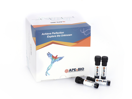
Live-Dead Cell Staining Kit
The Live-Dead Cell Staining Kit provides fluorescent markers to distinguish between live and dead cells in cultured cell populations. It uses two complementary dyes: Calcein-AM and Propidium Iodide (PI). Calcein-AM is a membrane-permeable non-fluorescent ester that can enter intact cells and is converted to Calcein inside the cell, emitting green fluorescence (excitation/emission: ~490/515 nm). PI is a membrane-impermeable nucleic acid dye that enters compromised cells and intercalates with cellular nuclear DNA, emitting red fluorescence (excitation/emission: ~535/617 nm). Applications include viability assays for flow cytometry, fluorescence microscopy, and drug cytotoxicity or apoptosis studies. Compared to single dye or Trypan Blue methods, the dual staining approach allows for the simultaneous visualization and counting of both live and dead cell populations.
An upgraded version of the Live/Dead Cell Staining Kit (K2247), including buffer, is also available.
- 1. Xiao-yan You, Xiang-yang Li, et al. "Development of NAFLD‐Specific Human Liver Organoid Models on a Microengineered Array Chip for Semaglutide Efficacy Evaluation." Cell Proliferation, 2025; 0:e70118
- 2. Tuo Deng, Jixing Li, et al. "A snail glycosaminoglycan-derived patch inspired by extracellular matrix accelerates diabetic wound healing via promoting re-epithelization." Volume 368, Part 2, 15 November 2025, 124168
- 3. Cuiping Sun, Zhiyong Du, et al. "Transglutaminase 2 nuclear localization enhances glioblastoma radiation resistance." Discov Oncol. 2025 May 30;16(1):952 PMID: 40445445
- 4. Mengmeng Jin, Yi Hou, et al. "Polydopamine-Coated Copper-Doped Mesoporous Silica/Gelatin–Waterborne Polyurethane Composite: A Multifunctional GBR Membrane Bone Defect Repair." J Funct Biomater. 2025 Apr 1;16(4):122 PMID: 40278230
- 5. Deyu Zhang, Songze Song, et al. "Glutamine binds HSC70 to transduce signals inhibiting IFN-β-mediated immunogenic cell death." Dev Cell. 2025 Mar 7:S1534-5807(25)00117-0 PMID: 40086433
- 6. Yu-Yao Li, Xiao-Hong Zhu, Zedong Cai. "Injectable Multifunctional Hemostatic Adhesive for the Hemostasis of Non‐Compressible Hemorrhage and Anti‐Infection of Bacterial Wounds." Macromol Biosci. 2025 Nov;25(11):e00294 PMID: 40605112
- 7. Hui Wang, Yue Zhu, et al. "Ginsenoside Rb1 improves human nonalcoholic fatty liver disease with liver organoids-on-a-chip." Engineered Regeneration 1 June 2024 Medicine, Engineering
- 8. Yeyang Wang, et al. "Electrospun silk fibroin/fibrin vascular scaffold with superior mechanical properties and biocompatibility for applications in tissue engineering." Sci Rep. 2024 Feb 16;14(1):3942 PMID: 38365964
- 9. Junyu Chen, et al. "Magnetic scaffold constructing by micro-injection for bone tissue engineering under static magnetic field." Journal of Materials Research and Technology. Volume 29, March–April 2024, Pages 3554-3565
- 10. Priyavrat Vashisth, Cameron L. Smith, et al. "Choline Carboxylic Acid Ionic Liquid-Stabilized Anisotropic Gold Nanoparticles for Photothermal Therapy." FORUM ARTICLE. January 29, 2024
- 11. Tanchen Ren, et al. "Enhancing aortic valve drug delivery with PAR2-targeting magnetic nano-cargoes for calcification alleviation." Nat Commun. 2024 Jan 16;15(1):557 PMID: 38228638
- 12. Chao Zhang, Hong Huang, et al. "DNA supramolecular hydrogel-enabled sustained delivery of metformin for relieving osteoarthritis." ACS Appl Mater Interfaces. 2023 Apr 5;15(13):16369-16379 PMID: 36945078
| 500 Tests | 1000 Tests | |
| Calcein-AM Solution (2 mM) | 50 μL | 50 μL × 2 |
| PI Solution (1.5 mM) | 150 μL | 150 μL × 2 |
Store at -20°C, protect from light. Calcein-AM is easily hydrolized by moisture. Keep it desiccate and tightly close the cap after the use. | ||


