JavaScript seems to be disabled in your browser. For the best experience on our site, be sure to turn on Javascript in your browser.
Tel: +1-832-696-8203
Email: [email protected]
Worldwide Distributors
In vitro transcription of capped mRNA with modified nucleotides and Poly(A) tail
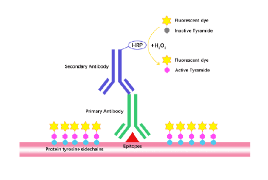
TSA (Tyramide Signal Amplification), used for signal amplification of ISH, IHC and IC etc.
Separation of phosphorylated and non-phosphorylated proteins without phospho-specific antibody
A convenient and sensitive way for cell proliferation assay and cytotoxicity assay
Protect the integrity of proteins from multiple proteases and phosphatases for different applications.
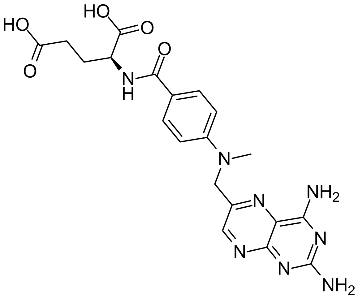
Methotrexate, a folate antagonist, is a potent anti-inflammatory agent when used weekly in low concentrations, the anti-phlogistic action of which is due to increased adenosine release at inflamed sites. Studies have demonstrated that methotrexate polyglutamates are active inhibitors of several enzymatic reactions, including dihydrofolate reductase. On consideration of biochemical pharmacology of methotrexate of methotrexate, it is taken up by cells and tissues and converted to methotrexate–polyglutamates, long-lived derivatives that retain biochemical and biologic activity within the cell. There is evidence that methotrexate does not act in rheumatoid arthritis (RA) simply as a cytotoxic agent for the cells responsible for the inflammation.
Reference
Bruce N. Cronstein, Dwight Naime, Edward Ostad. The antiinflammatory mechanism of methotrexate. Increased adenosine release at inflamed sites diminishes leukocyte accumulation in an in vivo model of inflammation. Journal of Clinical Investigation. 1993 December; 92(6): 2675–2682.
Bruce N. Cronstein. The mechanism of action of methotrexate. Rheumatic Disease Clinics of North America Volume 23, Issue 4, 1 November 1997, Pages 739–755.
Cell lines
activated T cells from human peripheral blood
Preparation method
The solubility of this compound in DMSO is >21.6 mg/mL. General tips for obtaining a higher concentration: Please warm the tube at 37 ℃ for 10 minutes and/or shake it in the ultrasonic bath for a while. Stock solution can be stored below -20℃ for several months.
Reacting condition
0.1-10 μM, 1-24 h
Applications
Methotrexate (0.1-10 μM) induced apoptosis of activated T cells from human peripheral blood. In PBL, treatment with MTX for 8 h induced apoptosis. Methotrexate at low (0.01 μM) and high (100 μM) concentrations inhibited cell proliferation without inducing apoptosis. Methotrexate-induced apoptosis required progression to the S phase of the cell cycle.
Animal models
Mice
Dosage form
Intraperitoneal injection, 2 mg/kg, once daily
Application
MTX exposure reduced thymus and spleen indices of mice. Methotrexate (≥5 mg/kg) markedly decreased white blood cells, thymic and splenic lymphocytes. Intraperitoneal injection with methotrexate for 3-4 wk increased splenocyte AICAR content, raised adenosine concentrations in exudates from carrageenan-inflamed air pouches, and markedly inhibited leukocyte accumulation in inflamed air pouches in mice.
Other notes
Please test the solubility of all compounds indoor, and the actual solubility may slightly differ with the theoretical value. This is caused by an experimental system error and it is normal.
References:
[1]. Genestier L, Paillot R, Fournel S, et al. Immunosuppressive properties of methotrexate: apoptosis and clonal deletion of activated peripheral T cells[J]. Journal of Clinical Investigation, 1998, 102(2): 322.
[2]. Gu S, Wu Y, Yang J. Screening of cytoprotectors against methotrexate-induced cytogenotoxicity from bioactive phytochemicals[J]. PeerJ, 2016, 4: e1983.
[3]. Cronstein B N, Naime D, Ostad E. The antiinflammatory mechanism of methotrexate. Increased adenosine release at inflamed sites diminishes leukocyte accumulation in an in vivo model of inflammation[J]. Journal of Clinical Investigation, 1993, 92(6): 2675.