JavaScript seems to be disabled in your browser. For the best experience on our site, be sure to turn on Javascript in your browser.
Tel: +1-832-696-8203
Email: [email protected]
Worldwide Distributors
In vitro transcription of capped mRNA with modified nucleotides and Poly(A) tail
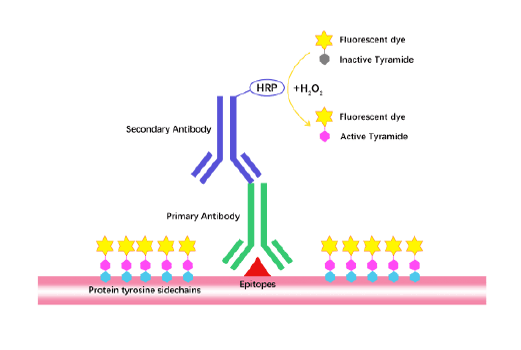
TSA (Tyramide Signal Amplification), used for signal amplification of ISH, IHC and IC etc.
Separation of phosphorylated and non-phosphorylated proteins without phospho-specific antibody
A convenient and sensitive way for cell proliferation assay and cytotoxicity assay
Protect the integrity of proteins from multiple proteases and phosphatases for different applications.
Epidermal Growth Factor (EGF) was originally discovered in crude preparations of nerve growth factor prepared from mouse submaxillary glands as an activity that induced early eyelid opening, incisor eruption, hair growth inhibition, and stunting of growth when injected into newborn mice. Human EGF was isolated from urine based on its inhibitory effect on gastric secretion and named urogastrone, accordingly. EGF is prototypic of a family of growth factors that are derived from membrane-anchored precursors. All members of this family are characterized by the presence of at least one EGF structural unit (defined by the presence of a conserved 6 cysteine motif that forms three disulfide bonds) in their extracellular domain. EGF is initially synthesized as a 130 kDa precursor transmembrane protein containing 9 EGF units. The mature soluble EGF sequence corresponds to the EGF unit located proximal to the transmembrane domain. The membrane EGF precursor is capable of binding to the EGF receptor and was reported to be biologically active. Mature human EGF shares 70 % a.a. sequence identity with mature mouse and rat EGF
Gene ID
1950
Accession #
P01133
Alternate Names
Source
Yeast
M.Wt
Approximately 6.0 kDa, a single glycosylated polypeptide chain containing 51 amino acids.
AA Sequence
NSDSECPLSH DGYCLHDGVC MYIEALDKYA CNCVVGYIGE RCQYRDLKWW E
Appearance
Sterile Filtered White lyophilized (freeze-dried) powder.
Stability & Storage
Use a manual defrost freezer and avoid repeated freeze-thaw cycles.
- 12 months from date of receipt, -20 to -70 °C as supplied.
- 1 month, 2 to 8 °C under sterile conditions after reconstitution.
- 3 months, -20 to -70 °C under sterile conditions after reconstitution.
Formulation
Lyophilized from a 0.2 μm filtered concentrated solution in PBS, pH 7.4.
Reconstitution
We recommend that this vial be briefly centrifuged prior to opening to bring the contents to the bottom. Reconstitute in sterile distilled water or aqueous buffer containing 0.1 % BSA to a concentration of 0.1-1.0 mg/mL. Stock solutions should be apportioned into working aliquots and stored at ≤ -20 °C. Further dilutions should be made in appropriate buffered solutions.
Biological Activity
Fully biologically active when compared to standard. The ED50 as determined by a cell proliferation assay using murine Balb/c 3T3 cells is less than 0.1 ng/ml, corresponding to a specific activity of > 1.0 × 10⁷ IU/mg.
Shipping Condition
Gel pack.
Handling
Centrifuge the vial prior to opening.
Usage
For Research Use Only! Not to be used in humans.