JavaScript seems to be disabled in your browser. For the best experience on our site, be sure to turn on Javascript in your browser.
Tel: +1-832-696-8203
Email: [email protected]
Worldwide Distributors
In vitro transcription of capped mRNA with modified nucleotides and Poly(A) tail
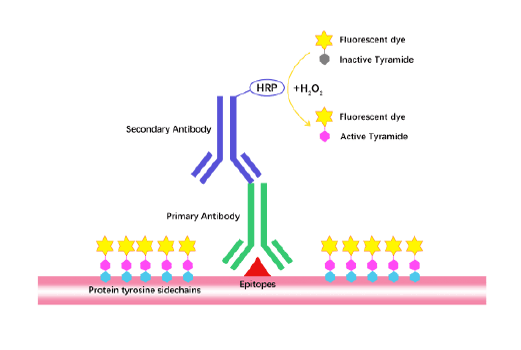
TSA (Tyramide Signal Amplification), used for signal amplification of ISH, IHC and IC etc.
Separation of phosphorylated and non-phosphorylated proteins without phospho-specific antibody
A convenient and sensitive way for cell proliferation assay and cytotoxicity assay
Protect the integrity of proteins from multiple proteases and phosphatases for different applications.
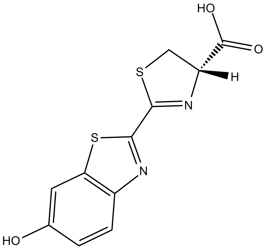
D-luciferin is a cell-permeable chemiluminescent luciferase substrate with a Km of approximately 2 μM. D-luciferin could emit lights upon oxidative decarboxylation in the presence of ATP. D-luciferin provides a bioluminescent signal for in vivo and in vitro detection of cellular ATP levels. D-Luciferin chould be used to assay the expression of the luciferase gene linked to a promoter of interest. Alternatively, D-luciferin and luciferase can be used to assess ATP availability in cellular or biochemical assays. D-luciferin could be administrated intravenously or intraperitonealy. In vivo and in vitro bioluminescence imaging (BLI) is a promising technique for non-invasive tumour imaging. The repeat ability coefficients of intravenously and intraperitonealy was 80.2% and 95.0%, respectively. PEmax of IP was 5.6 times higher for IV. When compared with IP, IV administration showed better repeatability and better sensitivity. It would be more beneficial to evaluate the accurate tumor burden of the small tumors rather than the larger tumors [1].
Reference:Keyaerts M, Verschueren J, Bos T J, et al. Dynamic bioluminescence imaging for quantitative tumour burden assessment using IV or IP administration of D-luciferin: effect on intensity, time kinetics and repeatability of photon emission[J]. European journal of nuclear medicine and molecular imaging, 2008, 35(5): 999-1007.